How Common Is Ovarian Dysgerminoma?
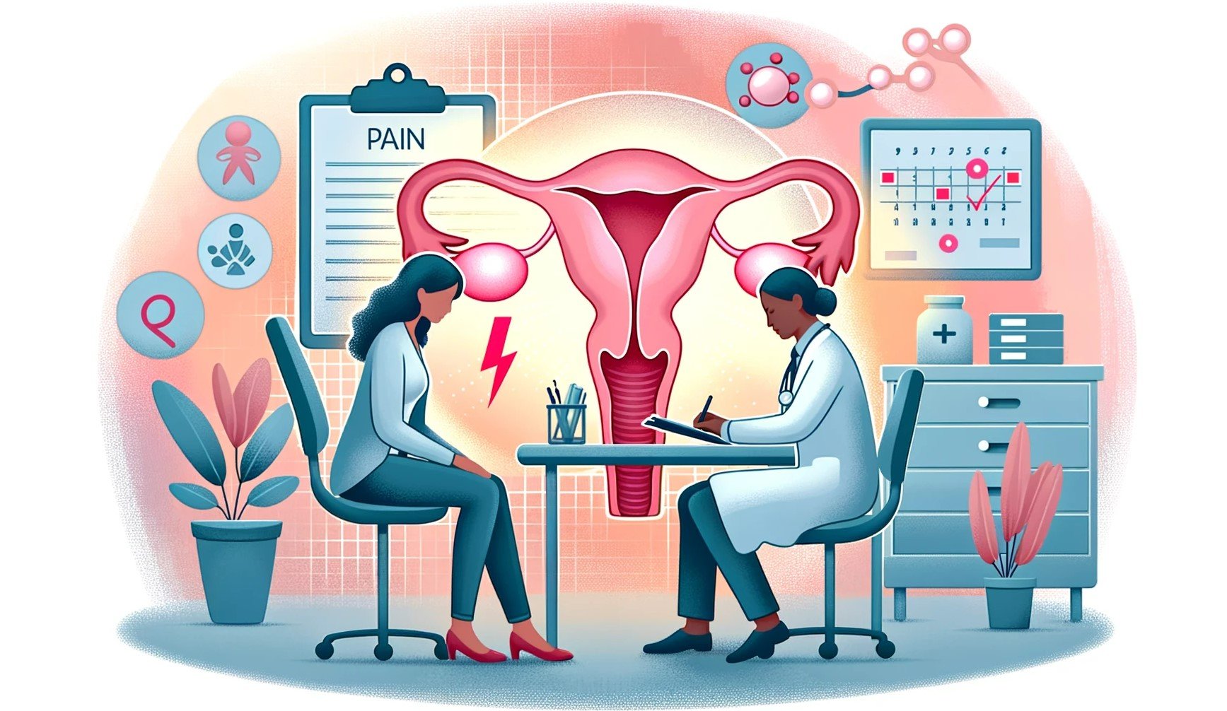
Ovarian dysgerminoma is a rare and specific type of ovarian tumour. It falls under germ cell tumours. It strikes women in their teens and early twenties. It represents only about 2% of the total cases. However, it demands attention. They're responsible for 32.8% of malignant ovarian germ cell tumours.
It accounts for a mere 1–5% of all ovarian cancers. These tumours occur during a woman's reproductive years. The incidence hovers between 2.8 and 11 cases per 100,000 pregnancies. Thus emphasizing the rarity of this condition.
Ovarian dysgerminomas are unique. Even though they're rare, they respond well to treatments like radiation and chemotherapy. This makes their outlook better compared to other ovarian cancers. Most people, about 90%, diagnosed it early in Stage I, when it hadn't spread. This gives it a strong chance of beating it.
The good news continues when looking at survival rates.
If diagnosed early, the 5-year survival rate is over 95%. And it rarely comes back, especially if found in just one ovary. Mostly, young women in their late teens to early 30s get it. It's a small portion of all ovarian cancers, only about 1–5%. But it's crucial to know about it and diagnose it early.
Some intriguing insights into ovarian dysgerminoma
What Are the Early Signs and Symptoms of Ovarian Dysgerminoma?
The early signs and symptoms of ovarian dysgerminoma can be somewhat nonspecific. This makes diagnosis challenging. Some of the early signs and symptoms are:
- Pelvic pain or discomfort
- Abdominal bloating.
- Swelling or a mass in the lower abdomen.
- Irregular menstrual periods.
- Nausea and vomiting

- Unexplained weight loss.
- Changes in urinary habits.
- Fatigue.
- Pain during sexual intercourse.
- Elevated levels of certain hormones
- Breast tenderness
- Abnormal hair growth.
Early diagnosis and treatment of ovarian dysgerminoma can lead to a good prognosis. Did you notice any such symptoms?
Don't worry, schedule your appointment today
What Are the Causes and Risk Factors of Ovarian Dysgerminoma?
Ovarian dysgerminoma is an uncommon ovarian tumour. Though rare, it's the top malignant germ cell tumor in the ovary. The exact reason why it develops is a mystery, but some things might raise the risk.
Causes: Experts still do not fully grasp why ovarian dysgerminoma happens. It's believed that DNA changes in ovarian cells might trigger it. These changes cause cells to grow wildly, forming tumors. The detailed genetic reasons behind it remain a puzzle.

Risk Factors:
Age: These tumors often appear in young women under 30.
Gonadal Dysgenesis: Women with this condition have ovaries that didn't grow right. It heightens the risk. Turner syndrome, a specific type of this condition, also increases the risk.
Swyer Syndrome: People with this have female looks outside but male organs inside that didn't grow right. This raises the chance of these tumors.
Family Ties: If your family has had germ cell tumors, your risk might be higher. But this link isn't proven yet.
Other Conditions: There's a hint that things like polycystic ovary syndrome (PCOS) might raise the risk. But it's not proven yet.
Even with these risk factors, it doesn't mean you'll get this tumor. Many with risks don't get it. And many who get it don't have clear risks. It's essential to stay aware and check any concerns with a doctor.
Don't stop scrolling.
The Road to recovery, what lies ahead.
How is ovarian dysgerminoma diagnosed?
The diagnosis of ovarian dysgerminoma involves the following:
Medical History: including any relevant symptoms, risk factors, and family history of cancer.
Physical Examination: Size and condition of the ovaries and any abnormalities
Imaging Studies
Ultrasonography: transvaginal ultrasound for size, location, and tumour characteristics.
Computed tomography (CT) / MRI: for the tumor's extent and any potential spread to nearby structures or lymph nodes.
Blood tests, tumour markers and biopsy are also carried out.
Early detection and diagnosis of ovarian dysgerminoma are crucial for effective treatment.
So what are you waiting for? If you notice any ovarian dysgerminoma symptom, connect with your doctor and get the screening tests done promptly!
What are the treatment options for ovarian dysgerminoma?
Treatment options for ovarian dysgerminoma may include:
- Surgery:
Oophorectomy: Surgical removal of the affected ovary (oophorectomy) is often done if the tumour is in one ovary and its early stages.
Bilateral Salpingo-Oophorectomy: in more advanced cases or on both ovaries, both ovaries and fallopian tubes were removed. It prevents recurrence or spread.
- Lymph Node Dissection: Done to check for the spread of cancer to nearby lymph nodes.
- Chemotherapy: For advanced or high-risk cases
- Fertility Preservation: In young women, fertility-sparing surgery is considered.
- Radiation Therapy: when the tumor is unresectable or if there's a recurrence.
Take the first step toward recovery by contacting us for your treatment.
What is the Prognosis and Survival Rate for Ovarian Dysgerminoma Patients?
The prognosis and survival rate are generally quite favorable when the diagnosed tumour is treated in its early stages. But depends on the stage at diagnosis and the extent of spread.
Here are some key points:
- Stage at diagnosis: Patients with stage I or stage II dysgerminomas have a very high survival rate, often exceeding 90-95%. In advanced stages (III and IV), the prognosis may be less favourable but is still relatively good compared to other ovarian cancers.
- Age at diagnosis: Dysgerminomas typically occur in young women and girls. Younger patients tend to have a better prognosis than older patients. Also, the tumour is often less aggressive in this age group.
- Tumor size and characteristics: The size of the tumor, rate of growth and the presence of specific markers can influence the prognosis. Smaller tumors and those with less aggressive features often have a better outcome.
- Treatment and response: Surgery to remove the affected ovary or ovaries
A study found that the overall 5-year and 10-year survival rates were 95.1% and 91.7%, respectively.
- Follow-Up Care: Regular follow-up appointments are essential to monitor for any signs of recurrence or complications. These may involve imaging studies (like CT scans or MRIs) and blood tests to assess tumor markers (such as LDH and AFP).
Early diagnosis and treatment of ovarian dysgerminoma leads to good prognosis and most patients can be cured.
Take charge of your health and your life. Contact us today!
Wondering if it will affect your fertility?
What Are the Long-term Implications of Ovarian Dysgerminoma?
The long-term implications of Ovarian Dysgerminoma are:
Infertility: Ovarian dysgerminoma commonly requires surgical removal of one or both ovaries, leading to infertility in affected individuals.
Regular Follow-Up Care: Long-term implications necessitate regular follow-up appointments to check for cancer recurrence and monitor the individual's overall health and well-being.
Hormonal Imbalances: It may disrupt hormonal balance in the body, potentially leading to symptoms such as irregular menstruation, hot flashes, and mood swings. These hormonal imbalances can persist long-term and may require management.
Emotional and Psychological Impact: Individuals may experience anxiety, depression, or fear of cancer recurrence, which can affect their quality of life.
Treatment Outcomes: Long-term implications can vary based on the stage of the cancer at diagnosis and the effectiveness of treatment. Early-stage, successfully treated cases. With minimally invasive surgery, there are fewer long-term complications than advanced or recurrent cases.
The treatment plans should be personalised according to your specific situation.
How Can Ovarian Dysgerminoma Impact Fertility and Pregnancy?
Ovarian dysgerminoma affects fertility in various ways. Here's a breakdown of the treatments and their impacts:
- Surgery: Removing one or both ovaries is often necessary. This can significantly change a woman's ability to have kids.
- Chemotherapy: This is a usual treatment. It can harm fertility either for a while or forever.
- Saving Fertility: There are ways to help. Doctors might suggest surgery that spares fertility. Some fertility preservation techniques, like freezing eggs and saving embryos and eggs in a frozen state, are available.
- Menstrual Return: About 9 months after treatment, many women get their periods back. This is a good sign they might be able to have kids.
- Natural Birth: Many women can still have kids after treatment. But the individual cases must be different.
- Pregnancy Care: If a survivor gets pregnant, doctors will watch closely. There might be health issues from the treatment that need attention.
If you're facing this condition and hope to have kids, discuss your options with a specialist. They can guide you on the best steps to take.
References:
https://www.ncbi.nlm.nih.gov/pmc/articles/PMC7983548/
https://journals.sagepub.com/doi/10.1177/8756479315599082
https://ovarianresearch.biomedcentral.com/articles/10.1186/s13048-020-00674-z


