Ready to take a deeper dive into the world of post-gallbladder removal issues?
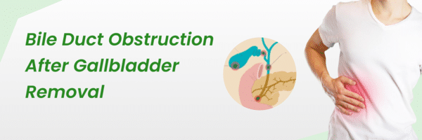
For some, the removal of the gallbladder isn't the end of the story. They face more challenges of bile duct stones post-surgery. These aren't remnants of the past; they have their own set of concerns.
Did you know it's reported that 10-15% of gallstone patients concomitantly suffer from bile duct stones? In fact, the incidence of recurrent bile duct stones, a common complication after gallstone surgery, ranges from 4% to 24%.
With new research and treatments emerging, there's hope for improved patient outcomes. Whether you're a post-op patient or a health enthusiast, stick with us.
Get ready to delve into the intricate world of bile duct stones - understanding their causes, symptoms, treatments, and cutting-edge advancements in medical science.
Take the first step toward healing. Request a Free Consultation.
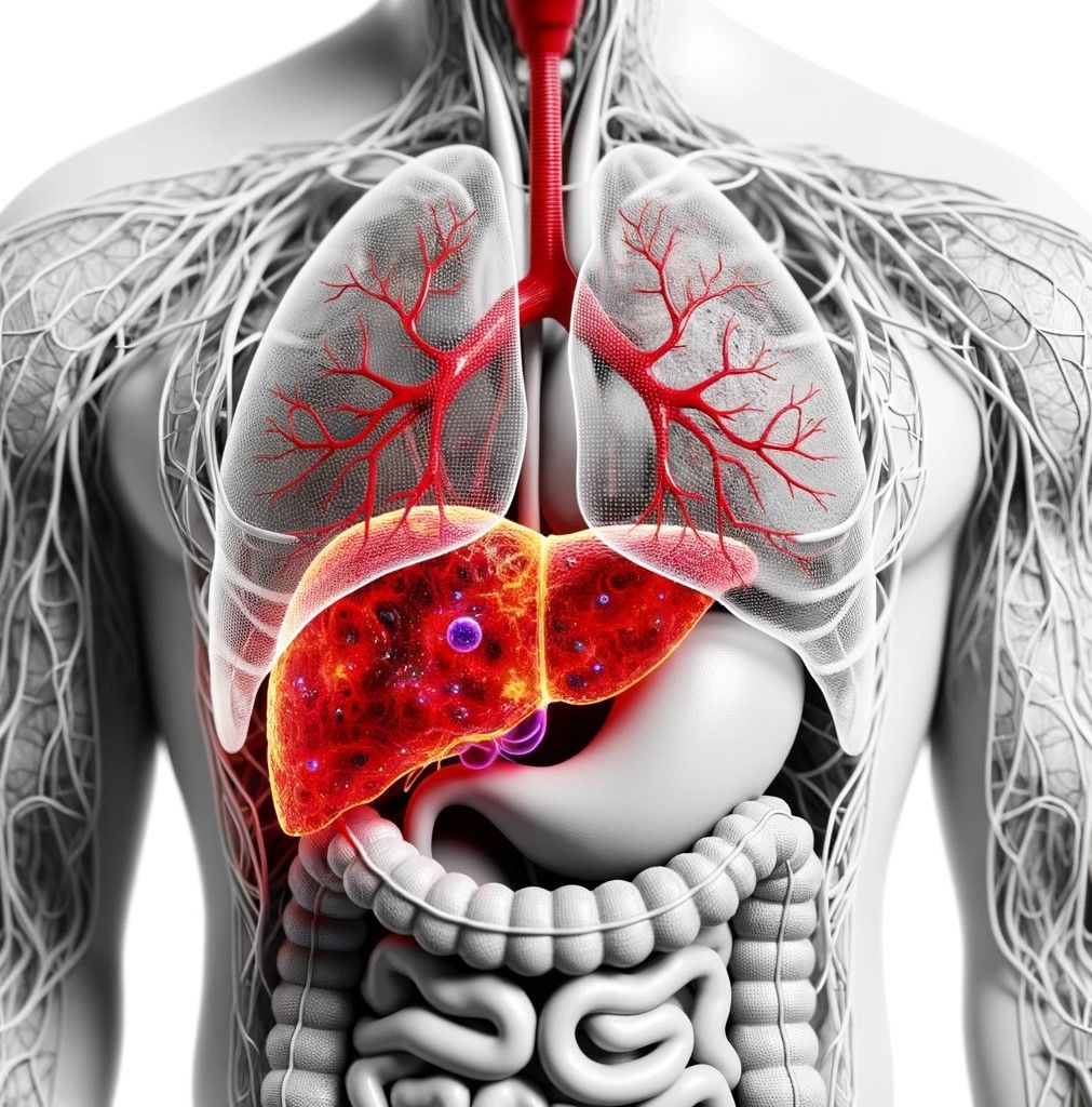
What causes bile duct stones to form after gallbladder removal?
Bile's Role: Did you know that the balance within your bile can play a crucial part in stone formation? Let’s dive into the contents of bile - it's a mixture of cholesterol, bilirubin, and bile salts. When there's an excess of cholesterol or bilirubin, stones are more likely to form.
Migration Matters: Surgery can act as a catalyst. During or after the procedure, stones might find their way from the gallbladder into the common bile duct. This migration is a common source of post-surgery stones.
Gallstones are commonly found in patients, affecting approximately 3.4% to 12% of individuals. It is common for gallstones to move from the gallbladder to the common bile duct, leading to the formation of bile duct stones among these individuals.
The Slow Flow Issue: Slow-moving bile can lead to stone formation. Especially if there's a narrowing in the bile duct or scarring from previous surgeries.
The Bacterial Connection: Beware of infections! Certain infections like E. coli or liver flukes might alter your bile composition and subsequently increase stone risk.
The Gallbladder's Role: Post-removal, bile moves directly from the liver to the intestine, changing its flow dynamics. This could potentially lead to stones.
Unique Anatomy: Anatomical differences in the bile duct system can lead to slower bile flow and stone formation.
Health Conditions: Certain health conditions may increase bilirubin production, which leads to stone formation.
Diet and Lifestyle: Your diet and lifestyle could be players in this game too. There is still ongoing debate about the complete influence of a high-fat diet, rapid weight loss, or a low-fiber diet. Besides, these factors are believed to have significant implications. Individuals over the age of 60 years are more likely to experience bile duct stones. Especially if they have obesity, a high-fat diet, or a sedentary lifestyle.
Did you know age affects the chance of getting bile duct stones with gallstones? Especially if you're over 60. The risk is much higher. For those under 60, there's an 8-15% chance. Moreover, elderly people tend to experience recurring bile duct stones more often than younger individuals.
But How Do These Stones Feel? Let's read ahead to learn more.
What are the symptoms of bile duct stones after gallbladder removal?

Experiencing pain in the upper right abdomen? Or maybe a yellow tinge in the eyes? These could be tell-tale signs of bile duct stones:
Pain: This is usually in the upper right abdomen and might spread to the shoulder or back.
Jaundice: A yellowing spectacle of your skin or eyes could be sounding the alarm for a blockage halting bile flow.
Urine Color: Dark urine and pale stools are potential indicators.
Nausea and Vomiting: These can be symptoms of blockage or inflammation.
Itchy Skin: Caused by increased bile salts in the bloodstream.
More severe symptoms like high fever and intense abdominal pain signal emergencies like cholangitis or pancreatitis.
Are you facing any of the above symptoms?
Don't delay— schedule your appointment today and get checked promptly!
How common are bile duct stones after gallbladder removal?
Post-gallbladder removal, bile duct stones aren't a rare occurrence. They could be:
1. Residual Stones: Missed during surgery or migrated during or post-surgery. This can occur due to the following reasons:
Migration from gallbladder or cystic duct post-surgery.
Challenges in detecting small stones with imaging.
2. New Stone Formation: Due to changes in bile composition or flow, new stones might develop.
Research estimates that 0.3% to 3% of patients might have residual stones post-surgery. While the risk for new stones is generally low, factors like time since surgery and age can influence it.
Are there any risk factors for developing bile duct stones after gallbladder removal?
Certain factors can heighten your risk:
Residual Stones: Missed or migrated stones from the original surgery.
Bile Duct Anatomy & Scarring: Variations or scarring can lead to stone formation.
Bacterial and Parasitic Infections: E. coli and liver flukes are key culprits.
Other Health Conditions: Hemolytic disorders, liver cirrhosis, and pregnancy can increase the risk.
Diet, Weight, and Genetics: Rapid weight loss, high-cholesterol diets, and even genetics play a role.
Are you concerned about developing bile duct stones after gallbladder removal? Let's dive into this topic!
Risk Factors: What Increases Your Chances?
Post gallbladder removal, several factors can bump up your risk of bile duct stones:
Residual Stones: Leftover stones from surgery might cause problems later.
Bile Duct Anatomy: Unusual structures can make stone formation easier.
Biliary Strictures: A narrowed bile duct can lead to stones.
Bacterial Infections: Some infections change bile composition, aiding stone formation.
Hemolytic Disorders: These can make stone formation more likely.
Liver Cirrhosis, Age, Pregnancy, Rapid Weight Loss, TPN, Diet, and Genetics: Each of these can play a role too. Keep in mind that having risk factors does not necessarily mean that you will experience complications. Regular medical check-ups and symptom awareness are your best defense.
Take charge of your health and your life. Contact us today!
How are bile duct stones diagnosed after gallbladder removal?
There are various imaging and procedural methods available to diagnose bile duct stones. Especially in people who have had their gallbladders removed and are experiencing symptoms of such stones. Here are the common diagnostic methods:
Ultrasound: This is usually the first-line imaging study. It's non-invasive and can often identify stones in the common bile duct (CBD). It can also detect dilatation of the bile ducts, which can occur if there's an obstruction.
MRCP: It provides detailed images of the bile ducts and pancreatic ducts using MRI. It's non-invasive and particularly useful for visualizing the bile ducts and detecting stones, strictures, or other abnormalities.
Endoscopic Ultrasound: This procedure uses a special endoscope equipped with an ultrasound device. EUS has the ability to detect smaller stones that may go unnoticed on other imaging modalities.
ERCP: ERCP is both a diagnostic and therapeutic procedure.
CT Scan: CT scan can detect bile duct stones, especially if they are large or if there's inflammation. It's more commonly used to rule out other differential diagnoses.
Liver Function Tests: Elevated liver function tests can indicate bile duct obstruction.
HIDA Scan: Evaluates bile flow from the liver to the intestine. Though it's not as commonly used for direct visualization of stones, it can help identify obstructions in the bile ducts.
Multiple tests can be used to diagnose bile duct stones after cholecystectomy. Timely evaluation and management are important to prevent complications.
Is surgery necessary to remove bile duct stones after gallbladder removal?
Good news: Not always! Surgery is not always necessary to remove bile duct stones after gallbladder removal. In fact, the most common method for removing bile duct stones after gallbladder removal is a non-surgical procedure called ERCP.
Let’s check ahead at all the available options:
- ERCP Steps:
- Insertion of an endoscope through the mouth to the duodenum.
- Catheter is introduced via the endoscope into the bile duct.
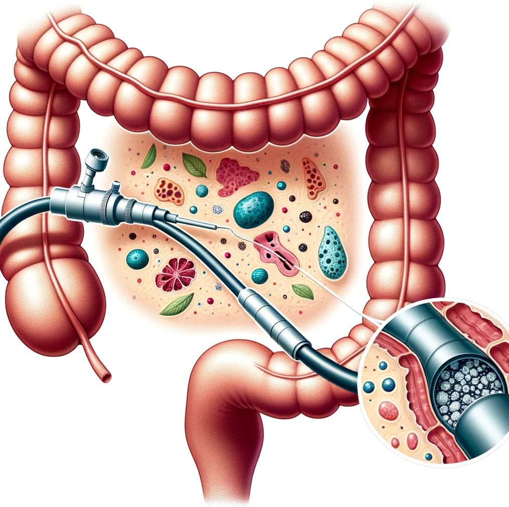
- Injection of contrast dye followed by X-ray imaging to see bile ducts and stones.
- Use of special instruments to manage stones.
- Potential sphincterotomy for easier stone removal.
- Temporary stent placement in challenging cases; removable later.
- Percutaneous Transhepatic Cholangiography (PTC):
- Alternative when ERCP is ineffective or not an option.
- Radiologist accesses bile ducts through the skin and liver.
- Allows for stone removal and dilation of strictures.
- Temporary drain or stent may be placed for optimal bile flow.
- Extracorporeal Shock Wave Lithotripsy (ESWL):
- Uses shock waves to fragment large stones.
- Broken-down stones pass naturally or can be extracted via ERCP.
- Medication:
- Ursodeoxycholic acid dissolves cholesterol-based stones.
- More common for gallstones.
- Not usually the first line for bile duct stones.
When is surgery the only option for the removal of bile duct stones after gallbladder?
If ERCP does not succeed or is not feasible, the doctor may recommend a surgical procedure called Open or Laparoscopic Common Bile Duct Exploration as an alternative. This method allows direct access to the bile duct and aids in its effective clearance. This can be done using traditional open surgery or minimally invasive laparoscopic techniques.
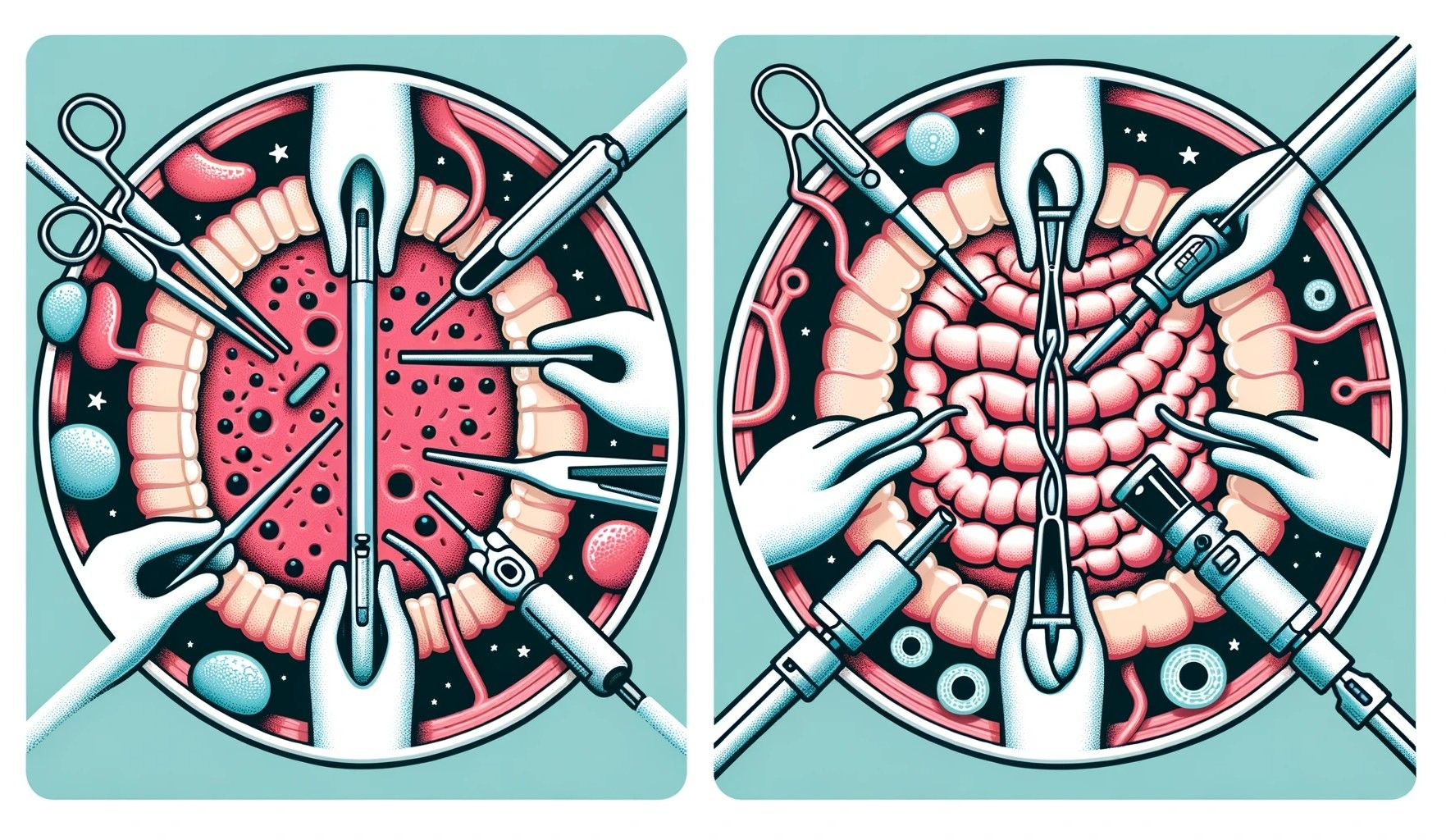
In certain situations, following the surgical clearance of the bile duct, the placement of a T-tube can be helpful. As it facilitates the external drainage of bile for a specific duration. This tube will be removed after a few days to weeks.
Book your appointment today for the best treatment for your unique situation.
Are there any complications associated with bile duct stones after gallbladder removal?
Yes, bile duct stones after cholecystectomy can lead to various complications. Here are some potential complications associated with bile duct stones after gallbladder removal:
- Acute Cholangitis: Imagine a stone acting as a roadblock in your bile duct, leading to the development of Acute Cholangitis, an infection that demands attention. The symptoms are - fever, jaundice, and right upper abdominal pain. If these are left unattended, it can escalate to septic shock.
Sounds scary, right? Timely treatment is your shield against this condition.
- Acute Pancreatitis: The bile duct and the pancreatic duct often share a common opening into the duodenum. When a stone gets stuck at this junction, it has the power to obstruct the flow of pancreatic enzymes This condition can ultimately result in pancreatitis - a painful inflammation of the pancreas. Symptoms include severe abdominal pain, nausea, vomiting, and elevated pancreatic enzymes in the blood.
- Biliary Cirrhosis: Chronic bile duct obstruction can lead to liver damage and cirrhosis over time.
- Bile Duct Injury: Persistent or large stones can injure the walls of the bile duct, leading to scarring, strictures (narrowing), or perforation.
- Bile Duct Strictures: Chronic inflammation from recurrent stones can lead to strictures or narrowing of the bile duct. It can cause obstructive jaundice and increase the risk of cholangitis.
- Gallstone Ileus: Though rare, a large stone can, in some cases, erode through the bile duct or gallbladder into the intestines and cause a blockage (ileus). This is more common with gallbladder stones but can happen with bile duct stones.
- Sepsis: If an infection like cholangitis is not treated promptly, it can spread to the bloodstream. This condition leads to sepsis, a severe and potentially fatal response to infection.
- Peritonitis: If a bile duct perforates or ruptures, bile can spill into the abdominal cavity, leading to inflammation known as peritonitis.
Keep your health in check. Regular check-ups and timely interventions are your best allies. Stay healthy, and stay informed!
Your well-being is our priority - call us to book your appointment today.
References:
https://www.ncbi.nlm.nih.gov/
https://www.sciencedirect.com/
https://journals.lww.com/ejos/fulltext/2023/42030/single_session_endoscopic_retrograde.5.aspx
https://onlinelibrary.wiley.com/doi/10.1002/deo2.294

