Overview
Did you know that, although a skin condition, basal cell carcinoma (BCC) can also occur rarely inside the mouth?
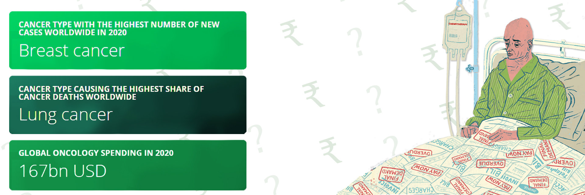
BCC is the most prevalent form of skin cancer, accounting for about 80% of all skin cancers. It usually appears as a shiny or pearly bump on sun-exposed areas of the skin. However, occurrences in the oral cavity are quite uncommon, representing less than 1% of all cases. This rarity makes it crucial to know how it manifests when it affects the mouth.
Understanding this unusual location for BCC is vital for early detection and effective treatment. Let's learn more about the signs to watch for and the importance of early diagnosis.
If you spot any persistent sores or unusual bumps in your mouth, schedule your appointment now for expert evaluation
Let's take a closer look at basal cell carcinoma
What is Basal Cell Carcinoma?
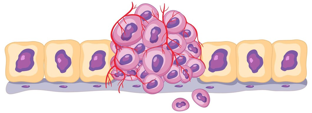
Did you know the skin has a defense layer deep inside?
Basal cell carcinoma (BCC) is a type of skin cancer that starts in these basal cells. This cancer is caused by too much sun exposure and appears on sun-exposed areas of the skin.
Here's how BCC usually looks:
- Appearance: It might appear as a small, shiny bump or a patch that seems pearly or light pink.
- Features: The bump can have tiny blood vessels visible or might have a sunken center.
But what happens when BCC shows up in a place you wouldn't expect, like the mouth?
Basal Cell Carcinoma in the Oral Cavity
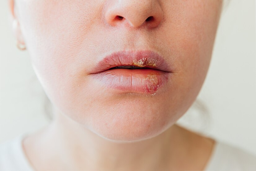
Inside the mouth, BCC is rare and looks different from the skin versions.
- Appearance: It might be a persistent white patch, a sore that doesn't heal, or an unusual growth that could bleed.
- Why It's Different: This type isn't caused by sunlight—since the inside of your mouth isn't exposed to the sun. The reasons it appears here are still not understood, which can complicate diagnosis.
What could lead to basal cell carcinoma in the oral cavity, especially when it's not exposed to the usual culprit, sunlight? Let’s explore some of the factors that might be involved.
Causes and Risk Factors

Genetic and Environmental Contributors
- Genetic Predisposition: Some people are more prone to developing basal cell carcinoma due to inherited conditions that affect skin cell growth and repair.
- Environmental Triggers: While UV light is a known risk factor for skin BCC, other environmental factors might contribute to oral BCC.
- Chronic Inflammation: Long-standing inflammation in the mouth, often from persistent infections or irritations (like fitting dentures), can increase the risk of cell changes leading to cancer.
- Immune System Function: A weakened immune system, which might be due to other diseases or medications, can also make it easier for cancers, including BCC, to develop.
Understanding these factors can help in the early identification and prevention of basal cell carcinoma in less common areas like the oral cavity.
Concerned about persistent mouth issues or risk factors mentioned here? Talk to us today.
Symptoms
Spotting the early signs of basal cell carcinoma (BCC) in the oral cavity can be tricky, especially since they can differ from those on the skin.
Early Warning Signs in the Oral Cavity:
- White or Red Patches: Unlike the pearly bumps often seen on the skin, BCC in the mouth may appear as flat white or red patches that persist.
- Non-Healing Sores: Sores that do not heal within a few weeks could be a sign of BCC.
- Bleeding: Any bleeding in the mouth without an obvious cause (like a cut from dental floss) should be evaluated.
- Lump or Thickening: A lump, bump, or increase in the thickness of the tissue inside your mouth that feels different from surrounding areas.
- Pain or Numbness: Unexplained pain or numbness in any area of the mouth or lips.
- Sensitivity: Oral lesions might not be as sensitive to touch as skin lesions, making them harder to detect based on feel.
Recognizing these symptoms early can lead to more timely and effective treatment.
Notice any of these symptoms in your mouth? It’s better to be safe and get them checked out. Contact us today!
Diagnosis and Staging
Diagnosis of Basal Cell Carcinoma in the Oral Cavity.
- Physical Examination: A doctor, often a dentist or a specialist, will start with a thorough examination of your mouth to check for any unusual signs like patches, sores, or lumps.
- Biopsy: If suspicious areas are found, a small sample of tissue will be taken (biopsy) and examined under a microscope to look for cancer cells.
- Imaging Tests: Depending on the biopsy results and the lesion's location, imaging tests like X-rays, CT scans, or MRIs might be used to see if the cancer has spread deeper into surrounding tissues.
Importance of Early Detection and Staging
- Better Outcomes: Early detection of BCC generally leads to simpler treatments with higher success rates.
- Staging: Understanding the cancer stage—how deep it has grown into the skin or if it has spread to other areas—helps doctors plan the best course of treatment.
Recognizing BCC early and understanding its stage can improve treatment effectiveness and patient prognosis.
Regular dental check-ups can catch early signs of problems, including BCC. If you have any concerns about changes in your mouth, don’t wait. Schedule your appointment now.
Treatment Options

If you’re facing basal cell carcinoma (BCC) in the oral cavity, understanding your treatment choices is key.
Treatment Methods:
- Surgical Options:
- Excision: This involves cutting out the tumor along with a bit of normal tissue around it to make sure all the cancer is removed.
- Mohs Surgery: This method removes the cancer layer by layer, checking each under a microscope during the procedure.
- Non-Surgical Options:
- Radiation Therapy: This can be a good choice if the tumor can’t be removed with surgery. It uses high-energy rays to kill cancer cells.
- Topical Treatments: Creams or gels that kill cancer cells might be used immediately for less severe BCC.
Advances in Treatment:
- Targeted Therapy: These treatments use drugs that attack specific parts of cancer cells. They are less likely to harm normal cells, making the treatment easier to handle.
- Immunotherapy: This approach boosts your immune system to help it recognize and destroy cancer cells, offering new hope for treating stubborn cancers.
Every treatment plan is customized, considering size of the tumor is, where it’s located, and how advanced the cancer is.
Dealing with BCC? A specialist can guide you through these options to find the best approach for you. Get in touch with us
Prevention Tips
Preventing basal cell carcinoma (BCC), especially in unusual places like the oral cavity, involves a mix of lifestyle adjustments and regular health checks.
Strategies to prevent BCC:
- Sun Protection: Shield your skin from UV rays with sunscreen, protective clothing, and shade, even though oral BCC isn't sun-related.
- Quit Smoking and Limit Alcohol: Tobacco and heavy drinking raise oral cancer risk. Quitting smoking and cutting back on alcohol can help.
- Oral Care: Keep your mouth healthy with regular brushing, flossing, and dental check-ups to reduce irritation and inflammation.
- Healthy Eating: Eat plenty of fruits and veggies for antioxidants that protect cells, including those in your mouth.
- Regular Check-Ups: Stay on top of medical and dental visits for early detection, especially if you've had BCC or other skin cancers.
Please note: By following these prevention tips, you can reduce your risk of developing BCC and maintain better health.
Conclusion
Living with basal cell carcinoma (BCC) requires resilience and adaptation. By making lifestyle adjustments and seeking support, you can confidently navigate this journey. Early detection is crucial, so stay proactive with self-examinations and medical check-ups.
Reference
https://www.ncbi.nlm.nih.gov/pmc/articles/PMC4126916/
https://pubmed.ncbi.nlm.nih.gov/11391100/
https://www.sciencedirect.com/science/article/abs/pii/S0002817714657524