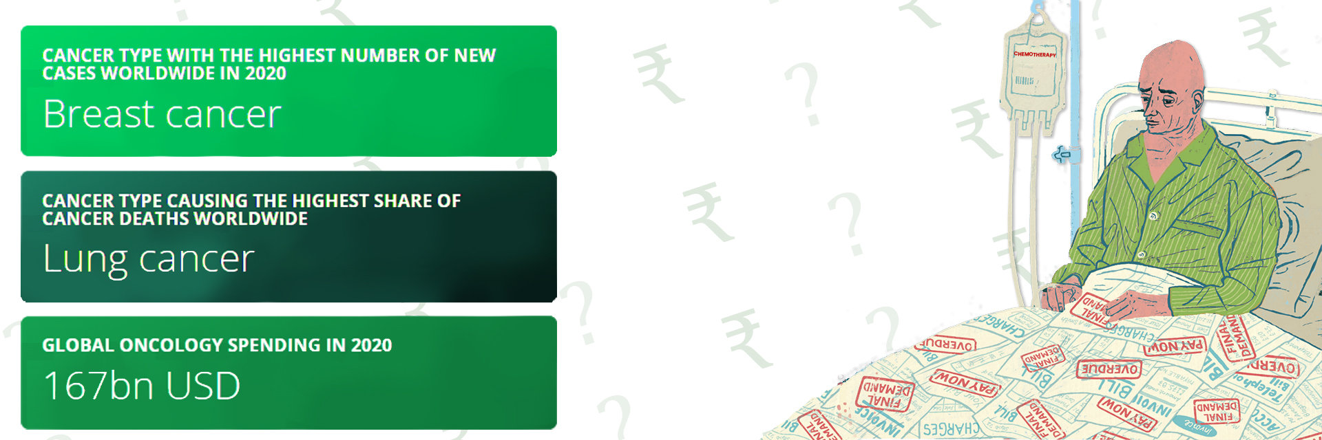
Overview
Wondering how lung cancer is treated? Let's understand.
A lobectomy, a common surgical method for treating lung cancer, involves removing one lobe of the lung that contains the cancer cells. This procedure is chosen when the cancer is localized to one area and hasn't spread to other parts of the lung, aiming to eradicate the disease.
But what if the cancer returns? In medical terms, "recurrence" refers to the cancer reappearing after it has been treated and eliminated. Recurrence after a lobectomy is not uncommon. Statistics show that approximately 30-55% of lung cancer patients may experience a recurrence, depending on factors like the stage of cancer at diagnosis and the completeness of the tumor removal.
Stay informed and proactive. It's vital for patients to understand the risks of recurrence and to keep regular appointments for monitoring.
Curious about how a lobectomy works? Let’s simplify it.
Understanding Lobectomy
The main goal is to eradicate the cancer from the lobe to prevent it from spreading.
What types of lobectomy are there?
- Traditional Lobectomy:
- Approach: Involves a larger incision in the chest.
- Purpose: Allows direct access to remove the affected lung lobe.
- Recovery: Generally longer due to the size of the incision.
- Invasive Lobectomy:
- Techniques: Includes video-assisted thoracic surgery (VATS) and robotic surgery.
- Approach: Uses smaller incisions, cameras, and specialized instruments.
- Benefits: Less post-operative pain and a quicker return to daily activities.
Causes of Recurrence After Lobectomy

After a lobectomy, which removes a cancer-affected lobe of the lung, there’s still a risk that lung cancer can recur. This means the cancer returns even after it seems to be completely removed.
Here are some common reasons and risk factors for recurrence:
- Microscopic Cancer Cells:
- Issue: Tiny cancer cells can remain undetected after surgery.
- Impact: These cells can grow over time, leading to recurrence.
- Quality of Surgical Margins:
- Issue: If cancer cells are close to the edges of the removed tissue, it suggests not all were removed.
- Impact: This increases the likelihood of cancer returning.
- Type and Stage of Cancer:
- Issue: Aggressive types and advanced stages of lung cancer are more prone to recur.
- Impact: These cancers are more challenging to cut completely.
- Patient’s Health and Genetics:
- Issue: Poor health and specific genetic factors can affect cancer behaviour.
- Impact: These can hinder recovery and ease the cancer's return.
- Smoking History:
- Issue: Continued smoking or a history of heavy smoking affects lung health.
- Impact: This can increase the risk of recurrence.
Stay proactive about your health. Understanding these risks is crucial for patients undergoing a lobectomy
Symptoms and Detection
Recurrence after lung cancer surgery can be subtle or pronounced, but recognizing the signs early can impact outcomes.
Common symptoms of recurrent lung cancer include:
- Persistent Coughing: A cough that doesn’t go away and worsens over time.
- Breathing Difficulties: Shortness of breath or wheezing, especially if new or worsening.
- Chest Pain: Pain in the chest area may be constant or occur only when coughing or breathing.
- Weight Loss: Unintended weight loss without trying.
- Bone Pain: New bone pain, such as the back or hips, may spread.
Diagnostic tools and tests for detecting recurrence:
- Imaging Tests:
- CT and PET Scans help visualize the lungs and detect tumor presence.
- MRI: Used especially to look for spread to the brain or spine.
- Biopsy:
- Collecting a small tissue sample from the lung or other areas where cancer might have spread to confirm recurrence.
- Blood Tests:
- Tests like the CEA test can track tumor markers that indicate cancer activity.
- Bronchoscopy:
- A tube with a camera is inserted into the airways to look for new tumors.
- Pulmonary Function Tests:
- Assess how well the lungs are working, such as obstruction or other issues from tumor growth.
Treatment Options for Recurrent Lung Cancer

Treatment for recurrent lung cancer varies based on factors like the cancer’s location, previous treatments, and the patient’s health.
Here’s an overview of treatment strategies for recurrent lung cancer:
- Surgery: If the recurrence is localized and the patient’s health allows, surgery may remove the new tumor.
- Radiation Therapy: This can target cancer cells in specific areas and is often used when surgery isn't viable.
- Chemotherapy: Useful for treating cancer that has spread and can be tailored based on the cancer's response to past treatments.
Exploring the latest in cancer treatment:
- Targeted Therapy:
- What It Does: Focuses on specific genetic mutations within cancer cells.
- Advantage: It can be more effective and less harmful to normal cells than traditional chemotherapy.
- Examples: Drugs like Erlotinib target the EGFR gene mutation, which is common in non-small cell lung cancer.
- Immunotherapy:
- What It Does: Boosts the body’s immune system to recognize better and attack cancer cells.
- Advantage: Offers a prolonged control of cancer, improving survival rates.
- Examples: Checkpoint inhibitors such as Nivolumab and Pembrolizumab have shown promise in treating advanced lung cancer.
Managing Treatment Side Effects
Treatment for recurrent lung cancer, while necessary, can lead to various side effects. Knowing what to expect and how to handle these can make the treatment journey more bearable.
Common side effects of recurrent lung cancer treatments include:
- Fatigue: Feeling tired is one of the most common effects of cancer treatment.
- Nausea and Vomiting: Often associated with chemotherapy and some targeted therapies.
- Hair Loss: A possible side effect of chemotherapy.
- Skin Reactions: Such as rash or itchiness, which is especially common with targeted therapies and radiation.
- Decreased Appetite: This leads to weight loss and decreased energy.
- Mouth Sores: Caused by both chemotherapy and radiation.
- Increased Risk of Infections: Due to a weakened immune system from treatments like chemotherapy.
Tips and advice for managing these side effects:
- Stay Hydrated: Drink plenty of fluids to help reduce nausea and keep your body strong.
- Nutrition is Key: Eat small, frequent meals to boost energy and manage weight loss.
- Gentle Exercise: Walking can boost energy levels and combat fatigue.
- Skin Care Routine: Use mild soaps and moisturizers to soothe dry or irritated skin.
- Oral Care: Maintain good oral hygiene and use a soft-bristled toothbrush to manage mouth sores.
- Manage Stress: Techniques such as meditation, yoga, or therapy can support emotional health.
Keen to know what’s new in the fight against recurrent lung cancer? Let’s take a look.
Latest Research and Developments
Recent studies and clinical trials continue to push the boundaries of what’s possible in treating recurrent lung cancer, bringing new hope to patients.
Recent highlights in the field include:
- Clinical Trials: Ongoing trials are exploring combinations of immunotherapy and targeted therapy, which show potential in reducing recurrence rates and improving survival.
- Genetic Profiling: Genomic sequencing advances allow doctors better to understand the specific characteristics of a patient’s cancer, leading to more personalized treatment plans.
- Emerging Therapies: Therapies like CRISPR gene-editing and CAR T-cell therapy are being studied for their potential to target and kill cancer cells more.
Conclusion
Understanding the causes and risk factors can help in managing the condition. From surgical options to innovative treatments like immunotherapy, the approach to managing recurrent lung cancer is becoming more tailored and effective. Side effects can be challenging but are manageable with the right strategies and support.
There is always a way forward. To those dealing with lung cancer recurrence: know that medical science is evolving, bringing new treatments and hope. Staying informed, proactive in monitoring, and working with your healthcare team are crucial steps in navigating this journey.
Reference:
https://wjso.biomedcentral.com/articles/10.1186/s12957-021-02165-x
https://acsjournals.onlinelibrary.wiley.com/doi/10.1002/cncr.24625