Get answers for your health queries from top Doctors for FREE!

100% Privacy Protection

100% Privacy Protection

We maintain your privacy and data confidentiality.

Verified Doctors

Verified Doctors

All Doctors go through a stringent verification process.

Quick Response

Quick Response

All Doctors go through a stringent verification process.

Reduce Clinic Visits

Reduce Clinic Visits

Save your time and money from the hassle of visits.

Ask Free Question

  1. Home >
  2. Blogs >
  3. Why is bone marrow transplant done?
  • Cancer

Why is bone marrow transplant done?

By Vedant Jain| Last Updated at: 22nd Mar '24| 16 Min Read

Before we begin you need to understand what is bone marrow:

Bone marrow is the semi-solid sponge like tissues present in the center of most bones, primarily located in the ribs, vertebrae, breastbone, and bones of the pelvis. It includes stem cells that ultimately forms blood cells like white blood cells, red blood cells and platelets.

Stem produces RBC, WBC & Platelets

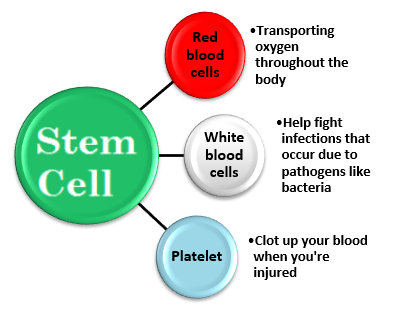
Bone marrow is where your hematopoietic stem cell are ultimately produced which help to make red blood cells, white blood cells and platelets.

The blood related diseases and cancers cause the cells to behave anti-socially.

This leads to abnormal behaviors such as un-controlled cell growth or breaking down of blood cells.

Treatment options used to treat these anomalies such as chemotherapy and radiation are given to you to kill all the damaged cells.

But these treatments attack both the good and bad cells.

Since stem cells contain immunity cells that help the body fight from infections and diseases, including cancer, it is important that they are replaced by a healthy bone marrow.

Usually, Bone marrow transplant is not the first line of treatment.

It is done when the bone marrow is unable to or not healthy enough to produce healthy cells and perform its functions due to chronic infections, disease or cancer treatments.

Hence the transplant is one of the last resorts for treating blood cancers and disorders prescribed by oncologist in India.

Cancers & Disorders that require Bone Marrow Transplants:

  1. Cancers that affect the marrow, such as Leukemia (unchecked growth of damaged blood cells), lymphoma (effects a type of white blood cells known as lymphocytes), and multiple myeloma (plasma cells become cancerous and grow out of control)
  2. Damaged bone marrow due to chemotherapy or radiation.
  3. Congenital neutropenia, which is an inherited disorder that causes recurring infections.
  4. Sickle cell anemia, which is an inherited blood disorder that causes red blood cells to misshapen and break down.
  5. Aplastic anemia, a rare condition in which the marrow stops making new blood cells.
  6. Thalassemia

Generally there are two ways that bone marrow transplant is done i.e. autologous and allogeneic. And both of these are used to treat different type of cancers. This can be classified as follows:
 

Autologous stem cell transplant:

Allogeneic Stem cell transplant:

  • Acute Myeloid leukaemia
  • Acute lymphoblastic leukaemia
  • Chronic myeloid leukemia
  • Myelodysplastic syndrome
  • Myeloproliferative disorders
  • Non-Hodgkins Lymphoma
  • Hodgkins Lymphoma
  • Chronic Lymphocytic leukemia
  • Multiple Myeloma
  • Aplastic anemia
  • Thalassemia
  • Fanconi’s anemia
  • Severe combined immunodeficiency
  • Paroxysmal Nocturnal Hemoglobinuria
  • Chronic myelomonocytic leukemia

Related Blogs

Question and Answers

. Heterogeneous Soft Tissue Nodule in the Right Lower Lobe (RLL) Size: 14 x 8 mm This nodule is described as heterogeneously enhancing, which suggests it may have varying levels of blood flow or different tissue densities within it. This could be indicative of a tumor. 2. Air Space Opacification in the Right Upper Lobe (RUL) Finding: There is patchy air space opacification with interlobular septal thickening in the posterior segment of RUL. This could represent infection, inflammation, or more concerningly, metastatic disease or lung cancer causing these changes. 3. Left-sided Pleural Effusion and Subsegmental Atelectasis Pleural Effusion: Mild left-sided pleural effusion is noted. Pleural effusion can occur in the context of metastatic disease or cancer. Atelectasis: This refers to partial lung collapse, which may occur when there is a mass obstructing the airflow or due to pleural fluid. 4. Enlarged Mediastinal and Hilar Lymph Nodes Lymphadenopathy: There are multiple enlarged and necrotic lymph nodes, most notably in the right hilar region, with the largest measuring 35 x 25 mm. Enlargement and necrosis of lymph nodes can be a sign of metastatic spread. The presence of enlarged lymph nodes in the mediastinum and hilum is typical of malignancy spreading beyond the primary lung site. 5. Liver Lesion Size: 14 x 13 mm lesion in the right hepatic lobe, which is well-defined and peripherally enhancing. A hypodense lesion could indicate a metastatic tumor, especially since it shows peripheral enhancement, a characteristic of some types of metastases. 6. Skeletal Lesions Multiple Lesions: There are mixed lytic and sclerotic bony lesions, some with soft tissue components. These lesions involve the vertebrae, ribs, glenoids, sternum, sacral ala, iliac bones, and femur. Soft Tissue Components: Some of the lesions, such as those in the ribs and iliac bones, have a soft tissue component, which suggests more advanced involvement, possibly indicating metastases. 7. Other Findings: No signs of emphysema, bronchiectasis, or pneumothorax were noted, which is reassuring as it reduces the likelihood of certain types of lung diseases. The liver, spleen, pancreas, kidneys, urinary bladder, and prostate all appear normal on imaging, which helps to rule out major issues in these organs. Impression: The findings of a heterogeneously enhancing solitary pulmonary nodule in the right lung, with associated hilar and mediastinal lymphadenopathy, along with a hepatic lesion and extensive skeletal involvement (with mixed lytic and sclerotic lesions), strongly raise concern for metastatic disease, most likely originating from the lung. The primary lung cancer is a potential consideration, though other primary sites are also possible. Next Steps: Histopathological correlation: This means a biopsy or tissue sample should be taken from one of the lesions (pulmonary, hepatic, or bone) to confirm whether the lesions are malignant and, if so, to identify the type of cancer. This will help determine the best course of treatment. The overall picture suggests a metastatic malignancy, likely of pulmonary origin, but further investigations and biopsy are essential to establish a definitive diagnosis and treatment plan.

Male | 58

Answered on 8th Mar '25

Read answer

Cancer Hospitals In Other Cities

Top Related Speciality Doctors In Other Cities

Cost Of Related Treatments In Country

Consult