What are the best foods to eat while on chemotherapy
Answered by Dr. Sandeep Nayak
It is important to eat a healthy, balanced diet during chemotherapy to keep your body functioning optimally. Foods that are mild in flavor, easy on your stomach, and nutrient-dense are some of the best options. Diet consisting of fruits vegetables and lot of fibers .

Surgical Oncologist
Questions & Answers on "Cancer" (374)
Related Blogs
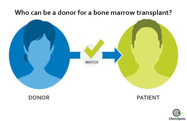
Who can be a donor for a bone marrow transplant in India?
Are you wondering who can be a donor for Bone Marrow Transplant in India? Then you are in the right place, below is the in-depth information about it.

Bone Marrow Transplant in India: Advanced Treatment Solutions
Discover advanced bone marrow transplant options in India. Trusted specialists, state-of-the-art facilities. Find hope and healing with personalized care.
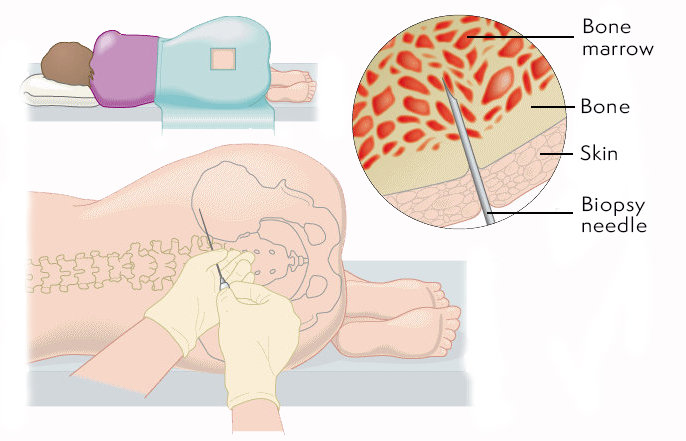
Risks and Complications of Bone Marrow Transplant in India
Here is the in-depth list of all the risks and complications involved in the bone marrow transplant.
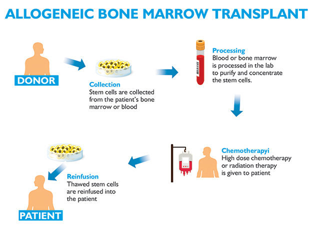
What is the Allogeneic Bone Marrow Transplant Cost in India?
Below is in-depth information and cost on Allogeneic Bone Marrow Transplant in India along with some of the best doctors to treat it.
Dr. Sandeep Nayak - Best Oncologist in Bangalore
Dr. Sandeep Nayak - Best oncologist in Bangalore. Experience of 19 years. Consults at Fortis, MACS & Ramakrishna. To book an appointment, call @ +91-98678 76979
Cost Of Related Treatments In Country
Top Different Category Hospitals In Country
Heart Hospitals in India
Prostate Cancer Treatment Hospitals in India
Kidney Transplant Hospitals in India
Cosmetic And Plastic Surgery Hospitals in India
Dermatology Hospitals in India
Endocrinology Hospitals in India
Gastroenterology Hospitals in India
Gynaecology Hospitals in India
Hematology Hospitals in India
Hepatology Hospitals in India
Top Doctors In Country By Specialty
Top Cancer Hospitals in Other Cities
Cancer Hospitals in Chandigarh
Cancer Hospitals in Delhi
Cancer Hospitals in Ahmedabad
Cancer Hospitals in Mysuru
Cancer Hospitals in Bhopal
Cancer Hospitals in Mumbai
Cancer Hospitals in Pune
Cancer Hospitals in Jaipur
Cancer Hospitals in Chennai
Cancer Hospitals in Hyderabad
Cancer Hospitals in Ghaziabad
Cancer Hospitals in Kanpur
Cancer Hospitals in Lucknow
Cancer Hospitals in Kolkata
- Home >
- Questions >
- What are the best foods to eat while on chemotherapy